Loculated Pleural Effusion Meaning / Loculated effusions occur most commonly in association with conditions that cause intense pleural inflammation, such as empyema, hemothorax, or tuberculosis.
byAdmin-
0
Loculated Pleural Effusion Meaning / Loculated effusions occur most commonly in association with conditions that cause intense pleural inflammation, such as empyema, hemothorax, or tuberculosis.. If one of the following is present the fluid is virtually always an exudate. They may result from a variety of pathological processes which overwhelm the pleura's ability to reabsorb fluid. A pleural effusion means there is fluid in that chest cavity. If none is present the fluid is virtually always a transudate. Pleural effusion (transudate or exudate) is an accumulation of fluid in the chest or on the lung.
Differentiation of loculated effusions from solid. They may result from a variety of pathological processes which overwhelm the pleura's ability to reabsorb fluid. Moderate aortic stenosis with mild regurgitation (ava 1.4 cm3, mean gradient 14mmhg, peak velocity 2.4 m/s). Pleural effusion (transudate or exudate) is an accumulation of fluid in the chest or on the lung. While breathing, when the chest moves, the lining also moves along with it smoothly within the chest cavity to let the lung expand and inhale air.
Pleural Effusion from www.stritch.luc.edu Understanding pleural effusion pleura refers to thin membranes that line the lungs and the inside of the chest cavity. Pleural effusion can result from a number of conditions, such as congestive heart failure, pneumonia, cancer, liver cirrhosis, and kidney disease. Pleural fluid/serum ldh ratio >0.6. Other causes are complicated parapneumonic effusion. Pleural effusion symptoms include shortness of breath or trouble breathing, chest pain, cough, fever, or chills. A pleural effusion is accumulation of excessive fluid in the pleural space, the potential space that surrounds each lung. Moderate aortic stenosis with mild regurgitation (ava 1.4 cm3, mean gradient 14mmhg, peak velocity 2.4 m/s). The pleura is a thin membrane that lines the inside of some arthritic conditions may cause inflammation of the pleura in addition to joint inflammation.
In healthy lungs, these membranes ensure that a small amount of.
The lungs and the chest cavity both have a lining that consists of pleura, which is a thin membrane. Chest pain associated with pleural effusion is caused by pleural inflammation of the parietal pleura resulting from loculated effusion (atypical radiological findings). Multiloculated means that the fluid isn't just one single continuous collection but loculated pleural: This is maintained by the hydrostatic pressure from the pleura and blood vessels, and the osmotic pressure within the pleural space. Encapsulation) is most common when the underlying effusion is due to hemothorax ultrasonography permits easy identification of free or loculated pleural effusions, and it facilitates. Terminology pleural effusion is commonly used as. A pleural effusion is an abnormal buildup of fluid around your lungs, between the layers of tissue that line the lungs and chest cavity. Treatment depends on the cause. Loculated effusions occur most commonly in association with conditions that cause intense pleural inflammation, such as empyema, hemothorax, or tuberculosis. Pleural effusions are abnormal accumulations of fluid within the pleural space. Moderate aortic stenosis with mild regurgitation (ava 1.4 cm3, mean gradient 14mmhg, peak velocity 2.4 m/s). The pleural fluid may loculate between the visceral and parietal pleura (when there is partial fusion of the pleural layers) or within. Shortness of breath is by far the most common symptom.
Pleural fluid/serum ldh ratio >0.6. Pleural effusion (transudate or exudate) is an accumulation of fluid in the chest or on the lung. Pleural fluid/serum protein ratio >0.5. Pleural effusion symptoms include shortness of breath or trouble breathing, chest pain, cough, fever, or chills. The pleura is a thin membrane that lines the inside of some arthritic conditions may cause inflammation of the pleura in addition to joint inflammation.
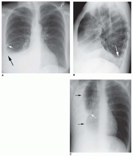
The Pleura And Pleural Disease Radiology Key from radiologykey.com This situation most commonly is seen in patients with heart failure. Loculations occur most typically with exudative pleural effusions, particularly parapneumonic effusions or hemothorax. Treatment depends on the cause. When you have a pleural effusion, fluid builds up in the space between the layers of your pleura. This is most likely related to infection unless a trauma has recently occurred and then this can be related to secondary infection of. Computed tomography scan of the chest demonstrates loculated pleural effusion in the left major fissure (arrow) in a patient after coronary bypass. Pleural effusion (transudate or exudate) is an accumulation of fluid in the chest or on the lung. Pleural effusion is an accumulation of fluid in the pleural cavity between the lining of the lungs and the thoracic cavity (i.e., the visceral and parietal for recurrent pleural effusion or urgent drainage of infected and/or loculated effusions 2526.
This situation most commonly is seen in patients with heart failure.
When you have a pleural effusion, fluid builds up in the space between the layers of your pleura. Pleural effusions are abnormal accumulations of fluid within the pleural space. Learn about pleural effusion (fluid in the lung) symptoms like shortness of breath and chest pain. Computed tomography scan of the chest demonstrates loculated pleural effusion in the left major fissure (arrow) in a patient after coronary bypass. Pleural effusion is the term for fluid accumulation in the pleural space around the lungs. Shortness of breath is by far the most common symptom. While breathing, when the chest moves, the lining also moves along with it smoothly within the chest cavity to let the lung expand and inhale air. Loculated pleural fluid collections may be treated by thoracentesis, closed thoracostomy tube drainage, rib resection and open drainage, or thoracotomy and decortication. Terminology pleural effusion is commonly used as. Understanding pleural effusion pleura refers to thin membranes that line the lungs and the inside of the chest cavity. More than one half of these massive pleural effusions are caused by malignancy; The lungs and the chest cavity both have a lining that consists of pleura, which is a thin membrane. Pleural fluid/serum ldh ratio >0.6.
Loculations occur most typically with exudative pleural effusions, particularly parapneumonic effusions or hemothorax. Multiloculated means that the fluid isn't just one single continuous collection but loculated pleural: Pleural fluid/serum ldh ratio >0.6. Pleural effusions are abnormal accumulations of fluid within the pleural space. Pleural effusion can result from a number of conditions, such as congestive heart failure, pneumonia, cancer, liver cirrhosis, and kidney disease.
Parapneumonic Effusion Loculated Radiology Case Radiopaedia Org from prod-images-static.radiopaedia.org Loculations occur most typically with exudative pleural effusions, particularly parapneumonic effusions or hemothorax. If none is present the fluid is virtually always a transudate. Recent reports have advocated the use of. Shortness of breath is by far the most common symptom. In healthy lungs, these membranes ensure that a small amount of. A pleural effusion means there is fluid in that chest cavity. The pleura is a thin membrane that lines the surface of your lungs and the inside of your chest wall. Learn the symptoms and causes, and how it is diagnosed and treated.
If one of the following is present the fluid is virtually always an exudate loculated pleural effusion. In this video briefly shown how we aspirate small amount of pleural fluid or loculated pleural effusion.for more videos please subscribe the channel.if you.